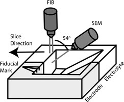
 Focused Ion Beam (FIB)/Scanning Electron Microscopy (SEM) is considered to be one of the most powerful techniques for three-dimensional morphological analysis and chemical composition characterization. It is widely used to study materials for energy applications and, coupled with computational analysis, it enables detailed 3D reconstructions of the examined sample. LiFePO4 (LFP) is an interesting cathode material for lithium-ion batteries because of its long cyclability and high safety. Because of the low ion and electron conductivity, LFP is always mixed with carbon black additives. Here we have used FIB/SEM tomography to study degradation in the carbon network in a laboratory-made LFP cathode. The SEM images were recorded at low accelerating voltage (1 kV) after ion milling. At such low voltage, primary electrons will not penetrate deep into the material so they will accumulate on the surface in regions with poor bulk electron conductivity, effectively changing the fraction of electrons entering the SEM detector and in turn making these regions appear relatively brighter. This principle has been used to study electron percolation in carbon structure. HRTEM studies were used to support of the low-kV FIB/SEM analysis of the carbon network degradation mechanism.
Degradation Studies on LiFePO4 Cathodes by FIB/SEM Tomography
Permanent link to this article: http://batteriselskab.dk/arrangementer/praesentationer/degradation-studies-on-lifepo4-cathodes-by-fibsem-tomography.htm